by Regina Rhoa, UoF Master Beekeeper
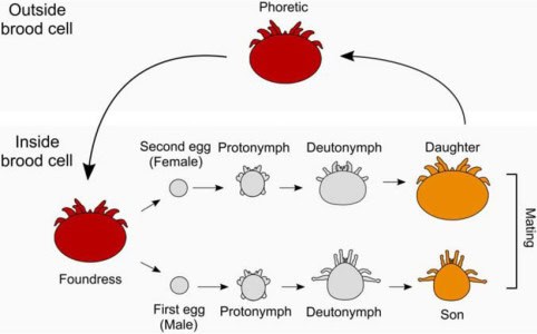
The life cycle of Varroa destructor mite consists of two main phases: the reproductive phase and the dispersal/feeding phase (formerly known as the phoretic phase). The term phoretic is defined as ‘riding’ on the host but not harming the host, but we now know that varroa mites feed on the adult honey bee.
During the onset of the reproductive phase, an adult female “foundress mite” enters a brood cell containing a 5th instar bee larva, which is the last larval stage before cell capping. Following entry, the foundress mite becomes temporarily immobilized in the brood food at the base of the cell, where she remains for up to six hours in worker cells and up to twenty hours in drone cells. Once the cell is capped, the developing bee larva emits an unidentified chemical signal (pheromone) that triggers egg production in the varroa mite.
Varroa mites, like the honey bee, utilizes a haploid-diploid sex determination system. In this system, females are diploid, possessing two sets of chromosomes inherited from both parents, while males are haploid, containing a single set of chromosomes from one parent, the mother. Consequently, male mites have only a maternal lineage, thus they have no father. The initial egg laid by the foundress is a male, typically deposited approximately 60-70 hours after cell capping. Subsequent eggs are females and are laid at intervals of 24–36 hours. Overall, a foundress mite produces up to five offspring within worker cells and six within drone cells. Over her lifetime, a foundress mite is capable of laying approximately 30 eggs, facilitating 4–5 brood cycles.

The foundress mite initiates the feeding site, which is predominantly located at the second abdominal sternite segment. While ground breaking work by Dr. Samuel Ramsey has shown that varroa mites feed on fat bodies of the adult honey bee (discussed more later), the varroa mite feeds on the hemolymph during the honey bee larval/pupal development, where the foundress mite creates a wound and injects digestive fluids.
This wound may result in structural harm to the developing larva/pupa, and varroa mites have the potential to transmit viruses, thereby compromising the bee's immune system. The mites suppress the pupal immune system, enabling viruses and bacteria to become more infectious.
Mating takes place between the son and all of the daughters. Interesting, the mating site is also the site of the fecal deposits, which is believed to be regulated by pheromones. The son begins by mating with the first daughter when she reaches maturity after her final molt, then continues with other successful daughters roughly every 30 hours. The male uses a pair of chelicerae—specialized appendages that form the main mouthparts—to transfer the spermatozoa (sperm).
The foundress mite uses these same mouthparts to pierce the honey bee host’s outer layer to feed during honey bee development as mentioned previously. Like honey bees, female varroa mites possess a spermatheca, which is a vascular organ designed to store sperm for future use, thus allowing the sperm to last for multiple brood cycles.
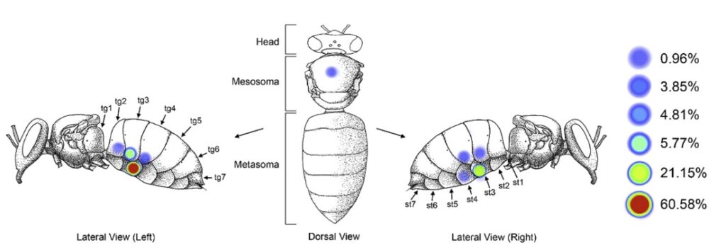
Because of the time required for mating with successive daughters, typically 1 daughter is successfully mated in worker cells, while 2 daughters are successfully mated in drone cells. Therefore, varroa mites demonstrate a preference for invading drone cells. Upon the adult bee's emergence from its cell, the foundress mite and her successfully mated daughters depart the cell, while the male and infertile daughters perish. At this point, the varroa mite enters the dispersal/feeding stage. Female mites must feed on adult honey bees, typically targeting nurse bees due to their enlarged fat bodies, which are essential for replenishing the mites' vitellogenin reserves—an important egg precursor. Dr. Samuel Ramsey has shown that varroa mites consume the fat bodies of the adult bees by embedding themselves beneath the bee's sternites (Fig. 3).
The majority hide under the third sternite, which is found on the underside of the bee (ventral side) and cannot be seen by beekeepers during normal inspections (see Fig. 4).
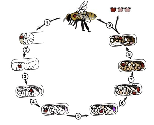
This feeding phase lasts approximately 4-6 days, after which the mite enters another brood cell to repeat the cycle (Fig. 5).
Varroa mites serve as vectors for numerous viruses found in honey bees, including Deformed Wing Virus (DWV A & B), Israeli Acute Bee Paralysis Virus (IABPV), and Acute Bee Paralysis Virus (ABPV). The transmission of these viruses by varroa mites significantly increases the viral effects due to the varroa piercing and damaging both the larval and adult bee tissue. Many viruses may remain asymptomatic unless viral loads reach exceedingly high levels, in-turn making detection by beekeepers challenging. Effective control of varroa mite populations is critical, as these mites play a major role in viral transmission and are a leading factor in honey bee colony mortality.
As varroa mite loads increase during the summertime, the production of drones conversely declines. This will shift the varroa mite to parasitize more worker cells. In addition, it is said that 90% of the varroa are hidden under the cell cappings, thus possibly shewing the results of mite washing. If at any time, you see a lot of capped brood, you can assume your mite infestation is much higher than your mite count, possibly facilitating treatment. Beekeepers should always look at all the factors going on in a colony when making mite management decisions, not just absolute numbers. It is important to do mite counts on a regular basis (generally monthly during active bee season). It is not only important to do mite counts before treatments, but after treatments to ensure your applied treatment worked.
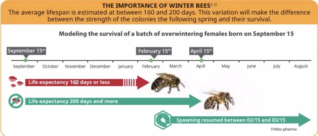
Varroa management becomes particularly crucial during late summer and early fall, as this period coincides with the formation of winter (diutinus or long-living) bees. These winter bees must survive for a longer period of time during which nurse bees rely on their enlarged fat bodies to produce brood food that feed larvae. Honey bee fat bodies are vital, multifunctional organs responsible for nutrient storage, detoxification, vitellogenin production (key for brood food and immunity), hormone regulation, and temperature maintenance. Newly emerged nurse bees possess robust fat bodies, which gradually diminish as they transition to house duties and eventually foraging. These fat bodies are essential for producing brood food, especially when pollen resources are scarce in winter. If varroa mites compromise the integrity of these fat bodies through feeding, the colony's ability to endure winter is diminished, resulting in reduced bee lifespan and potential colony decline. Beekeepers may assume their colonies are healthy as they approach winter; however, elevated viral loads and compromised bee health can result in mortality during early/mid-winter.
Honey bees typically have a lifespan of approximately 4–6 weeks from spring through early autumn. In contrast, winter bees must survive for six months or longer. As illustrated in Figure 6, colonies infested with varroa mites and their associated viruses will experience significant winter bee mortality by mid-winter, generally when beekeepers see colony losses. Conversely, if winter bees are free from varroa infestation, the colony population can remain robust until the queen resumes brood rearing in mid-winter to replenish any population losses. In the Tri-state, the queen will start laying in December and January but will ramp up in early March.
Egg Laying Overview
- After entering a capped brood cell, the female mite waits ~60–70 hours before laying her first egg (unfertilized male). Subsequent eggs (fertilized females) are laid every ~30 hours (up to 4–6 total).
- Foundress Mites: In subsequent cycles, varroa can lay the first egg as early as ~60 hours post-capping, due to retained reproductive readiness from prior cycles. They produce more viable offspring (potentially up to 2 viable daughters per worker cell) than in their first reproduction. Foundress mites can lay their first egg slightly earlier in subsequent cycles compared to daughter mites in their first reproduction leading to more viable offspring.
- Daughter Mites: Newly matured daughters typically wait ~70 hours to lay their first egg, as they need time to fully activate oogenesis (egg production) after emerging.
- Difference: The 10-hour gap is small but significant in synchronized brood cycles; foundress mites are more efficient, contributing to faster population buildup.
- Factors Influencing Timing: Cell type (drone cells allow the foundress to 2–3 extra eggs due to longer pupation); hive temperature (optimal 34–35°C); and mite health. This leads to 2 viable daughter offspring.
Virus Transmission Overview
- Foundress Mite Entry and Wound Creation
- The invading (foundress) female mite enters a capped brood cell ~12–24 hours before sealing.
- She pierces the pupa's integument (skin) to feed on hemolymph/fat body, creating a permanent feeding site/wound (often on the abdomen or thorax).
- If the foundress carries viruses (e.g., DWV, ABPV from previous feeding on adults), she injects them directly into the wound via saliva during feeding. This infects the pupa early.
- Daughter Mites Become Infected:
- The foundress lays eggs; offspring (protonymphs/deutonymphs) feed at the same wound site created by the mother.
- They acquire viruses horizontally from the infected pupal hemolymph (high viral titers at the wound).
- Result: Daughter mites emerge already carrying high viral loads, even if the mother had low levels.
- Dispersal and Further Spread:
- Mature daughter mites (now virus vectors) enter the dispersal/feeding phase on adult bees.
- When they invade new brood cells as foundresses, they repeat the cycle: create wounds → inject viruses → infect pupae → pass to next generation.
- This amplifies viruses exponentially—one infected foundress mite can seed dozens of new vectors.
- Key Viruses Involved (Replication)
- Deformed Wing Virus (DWV): Most common; wound feeding boosts titers 10⁶–10⁹-fold.
- Acute Bee Paralysis Virus (ABPV): Rapid activation via mite saliva.
- Others: KBV, IAPV, SBPV—similar wound-mediated spread.
The summary demonstrates that as the season advances, mites re-entering brood cells not only reproduce more rapidly but also transmit higher levels of viruses, resulting in increased mite populations and elevated viral loads. Delaying varroa management until later in the beekeeping season means contending with both substantial mite infestations and heightened viral presence. It is frequently reported by beekeepers that treatment was delayed until mid-to-late summer due to varrocide application restrictions; however, this approach can ultimately result in colony losses during winter, even if mite levels are reduced prior to the emergence of winter bees.
We as beekeepers don’t always have full control of where our honey bees forage for their nectar/pollen or get exposed to pesticides. We can give our bees a much better opportunity for survival by controlling varroa and their associated viruses. By better understanding varroa mite biology and the impact on our bees, we give our bees a fighting chance to survive until spring and hopefully produce a great honey crop or expand our apiaries.
References
- https://www.sciencedirect.com/science/article/pii/S0022519325003133
- https://www.nature.com/articles/s41598-023-49688-9
- https://www.ars.usda.gov/pacific-west-area/tucson-az/carl-hayden-bee-research-center/research/varroa/varroa-mite-life-cycle-and-reproduction/
- https://academic.oup.com/jinsectscience/article/22/1/18/6523143
- https://pubmed.ncbi.nlm.nih.gov/27209572/
- https://www.pnas.org/doi/abs/10.1073/pnas.1818371116
- https://www.nature.com/articles/s41467-024-44915-x
- https://extension.msstate.edu/publications/managing-varroa-mites-honey-bee-colonies
- https://academic.oup.com/jinsectscience/article/22/1/18/6523143
[Return to February 2026 Beeline newsletter]